Summary
Lameness diagnosis involves, visual gait assessment, and hands‑on examination to detect pain, swelling, or abnormal limb function. In horses, flexion tests, hoof testers, and advanced imaging such as X‑ray, ultrasound, scintigraphy, CT, or MRI may be used to pinpoint the source of pain. In dogs, gait observation, palpation, and radiographs are primary tools, with advanced imaging used for complex joint or bone issues. Treatment depends on the underlying cause and may include rest, NSAIDs, joint injections, corrective shoeing (for horses), physiotherapy, or surgical repair such as TPLO/TTA in dogs or tenotomy in horses. New technologies like inertial sensor systems and AI‑based gait analysis are improving early detection and monitoring of lameness, supporting more targeted therapy and better outcomes.
Explanation
Lameness is the number one cause of restricted mobility in horses, and can stem from a variety of conditions and injuries. We can pinpoint the underlying cause and create a treatment plan tailor-made for your horse’s activity level and performance expectations.
Lameness Exams
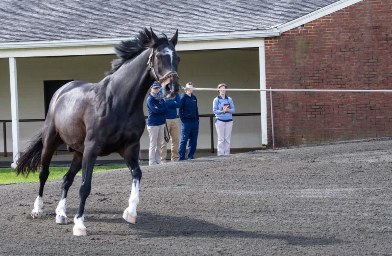
Your horse’s lameness exam will include a complete health history (if needed) and your input about the injury and onset of symptoms. We’ll perform a thorough physical examination, observe your horse in motion, and administer joint flexion tests. We’ll also apply hoof testers to look for pain or unusual levels of sensitivity in the feet.
We might also use nerve and joint blocks to numb portions of a limb. This helps us identify, through the process of elimination, the location of the pain. Occasionally, we might use x-rays or ultrasound for a proper diagnosis.
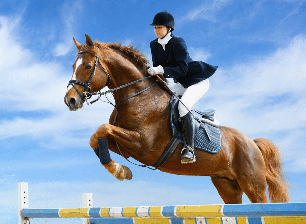
Physically immature horses that are subjected to repetitive stress on bones are prone to lameness. Immature bones may be anatomically normal but weak due to the age of the horse. Developmental orthopedic disease (such as limb deformities); poor conformation (such as pigeon toes or bowleggedness); improper hoof balance or shoeing; failure to adequately condition performance horses; improper footing (hard, slippery, or rocky surfaces); hard exercise; and repetitive stresses on bones, tendons, ligaments, and joints in performance horses can also cause lameness. An example of repetitive stress on bones is the continuous training of race horses around left-handed bends. This can produce shin soreness, stress fractures, or imbalances of the feet that abnormally distribute body weight among the limbs. Other factors that cause lameness include direct or indirect trauma to a limb, fatigue in racehorses racing over long distances, or inflammation—more often than not without infection—of joints, tendons, and ligaments.
Lameness in one part of a limb will often produce soreness in another area of the limb as well. It can also lead to a secondary lameness in the fore- or hindlimb on the opposite side of the body.
The Lameness Examination
A thorough investigation of a lame horse is necessary in order to ensure a correct diagnosis and appropriate treatment. The examination begins with a full medical history. The horse’s type, age, and training regimen may give important clues to the lameness. Your veterinarian will ask how much time has passed since the onset of lameness and how it has been managed. The length of time since the last shoeing will be noted, as well as any indication that the lameness improves with either rest or exercise. The horse’s response to anti-inflammatory or pain-relieving medications may provide useful information. Results of laboratory tests may reveal other problems that influence overall performance.
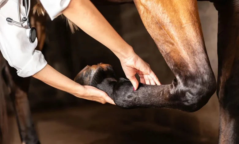
A detailed visual inspection is followed by a hands-on examination of the limbs in weight-bearing and non-weight-bearing positions to identify any heat, pain, swelling of joints, or abnormal tissue tension. Your veterinarian will also study your horse’s reactions, look for any muscle loss, and measure the range of movement in the joints. Your veterinarian will observe whether lameness seems to increase or decrease after flexing or extending the joint.
The feet are thoroughly examined, including the use of hoof testers to look for sore spots in the sole. Wear patterns of shoes and feet are noted. In addition to the legs and feet, the back and neck should be thoroughly examined with the horse restrained and standing on a level surface.
Diagnosis:
- History and Physical Examination:A veterinarian will begin by gathering information about the horse’s history and then perform a thorough physical exam, observing the horse’s gait, posture, and overall condition.
- Flexion Tests:These tests involve flexing specific joints to assess for pain or range of motion limitations.
- Hoof Testers:These tools help pinpoint areas of sensitivity in the hoof.
- Diagnostic Nerve Blocks:These are used to numb specific areas of the limb, helping to pinpoint the source of pain.
- Diagnostic Imaging:Radiographs (X-rays), ultrasound, or advanced imaging like MRI or CT scans may be used to visualize bones, joints, and soft tissues, according to Equine Sports Medicine.
- Arthroscopy:This minimally invasive surgical procedure allows for direct visualization of the joint and can be used to diagnose and treat certain conditions.
Treatment:
- Rest:Rest is often a crucial part of the treatment plan, and the duration of rest can vary depending on the injury.
- Rehabilitation:Physical therapy and controlled exercise can help restore range of motion and strength.
- Therapeutic Sheoing:Corrective trimming and shoeing can address hoof imbalances and reduce stress on certain areas of the limb.
- Surgery:In some cases, surgery may be necessary to repair damaged tissues or correct structural problems.
FAQs on Horse Lameness Diagnosis and Treatment
- What is lameness in horses?
It is an abnormal gait or movement due to pain, injury, or mechanical problems in a limb. - How is lameness first detected?
By observing the horse at rest and in motion (walking and trotting) for irregular movement. - What are common causes of lameness in horses?
Hoof abscesses, laminitis, tendon or ligament injuries, joint disease, and fractures. - What is the purpose of a flexion test?
To temporarily stress a joint and see if lameness increases, helping locate pain. - What tools help in diagnosing lameness?
Palpation, hoof testers, nerve blocks, radiographs, ultrasound, CT, or MRI. - Why are nerve blocks used?
To numb specific areas and find the exact source of pain by observing gait changes. - What role do radiographs play?
They help identify bone or joint abnormalities such as fractures or arthritis. - When is ultrasound preferred?
For evaluating soft tissue injuries like tendon or ligament damage. - What medical treatments are available?
Rest, NSAIDs, joint injections, shockwave therapy, and corrective shoeing. - What surgical treatments exist?
Procedures such as tenotomy, desmotomy, or tendon splitting for specific.
Need Veterinary Help?
Have questions about animal health, management, diseases, or treatments?
Our professional veterinary team is ready to assist you!
Consult Online: Contact Us
Email: professionaldvm129@gmail.com
Visit: www.veterinaryhub.info


aapq1n
pvj6kj