
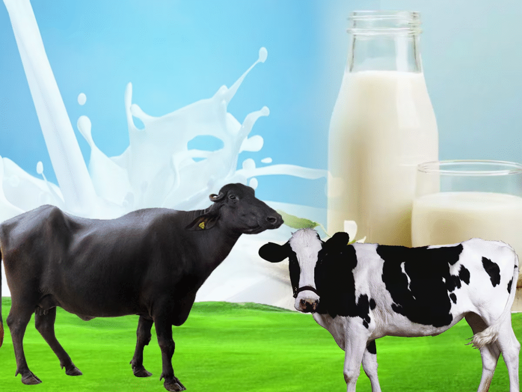
In this post let us go over the difference between Buffalo milk and Cow milk, look at the nutritional and mineral content, and decide which is more suitable for children and/or adults.
Buffalo milk is more thicker and contains slightly more vitamins and minerals than cow milk. However, cow milk more preferred due to following reasons:
- The fat and protein of cow milk are more easily digestible than those of buffalo milk.
- The cow milk has also got greater amount of vitamins and minerals.
- Buffalo milk is injurious to the development of children and only cow milk is useful to them (in the absence of mother’s milk. The buffalo milk is rich in fat, which children can’t digest. Because of indigestion they suffer from diarrhea. The fats in cow milk and buffalo milk differ from each other in their digestive properties. The percentage of volatile and soluble acids is greater in cow ghee, and consequently it is more easily digested.
- Cow milk, being easily digestible, is more beneficial to patients than buffalo milk.
- Cow milk is useful for intellectual growth of children.
Let us have closer look at nutritional value of both Buffalo and Cow milk.
Nutritional Chart
| Nutrition | Buffalo Milk | Cow Milk |
|---|---|---|
| Water | 81.1% | 87.8% |
| Protein | 4.5g | 3.2g |
| Fat | 8g | 3.9g |
| Carbohydrate | 4.9g | 4.8g |
| Energy | 110kcal | 66kcal |
| Sugar lactose | 4.9g | 4.9g |
| Saturated Fat | 4.2g | 2.4g |
| Monounsaturated Fat | 1.7g | 1.1g |
| Polyunsaturated Fat | 0.2g | 0.1g |
| Cholesterol | 8mg | 14mgg |
| Calcium | 195micg | 120micg |
As you can see from above chart buffalo milk has less water content and hence thicker than cow milk. It has also got higher protein and calcium and less cholesterol than cow milk. However, its high degree of fat and calories makes it less attractive. So, if you drink buffalo milk you really need to work out. Probably this is the reason why people who work hard in the farm preferred buffalo milk over cow milk.
Mineral Content
| Minerals | Buffalo Milk | Cow Milk |
|---|---|---|
| Calcium | 0.18% | 0.12% |
| Phosphorous | 0.14% | 0.10% |
| Magnesium | 0.02% | 0.01% |
| Sodium | 0.04% | 0.05% |
| Potassium | 0.11% | 0.15% |
| Chloride | 0.07% | 0.10% |
| Citrate | 0.18% | 0.18% |
There isn’t significant difference in the mineral content except for more calcium in buffalo milk and more potassium in cow milk.
Overall Comparison of Buffalo Milk and Cow
| Buffalo Milk | Cow Milk |
|---|---|
| Milk good for adults | Milk good for infant and adults |
| Milk is thick | Milk is thin |
| Milk is white | Milk is golden yellow |
| Milk is not easily digestible | Milk is very easily digestible |
| Milk has less cholesterol | Milk has more cholesterol |
| Milk has more fat | Milk has less fat. |
| Milk has more calories | Milk has fewer calories |
| 100 calories are derived for 100g buffalo’s milk | 70 calories are derived for 100 g buffalo’s milk |
| Milk has less water | Milk has more water |
| Milk has more protein | Milk has little less protein |
| Milk has slightly more carbohydrates | Milk has slightly less carbohydrates. |
| Milk has more saturated fatty acid, mono unsaturated fatty acid and poly unsaturated fatty acid | Milk has less saturated fatty acid, mono unsaturated fatty acid and poly unsaturated fatty acid |
| Milk has more calcium, iron, phosphorus etc. | Milk has less calcium, iron, phosphorus etc. |
| Milk has more Vitamin A | Milk has fewer Vitamin A but good in Vitamin E |
| Probably milk has less sulphur | Probably milk has more sulphur – good for active brain |
| Top three producers are India, Pakistan and China | Top three producers are Finland, Sweden and Ireland |
| Milk can be preserved for longer time naturally | Milk can’t be preserved for longer time naturally |
| A good buffalo gives 10 litres milk per day | A good cow gives 20 litres milk per day |
| Raw milk contains more pathogenic micro organisms | Raw milk contains less pathogenic micro organisms |
| Milk has lesser iodine | Milk has more iodine |
| Milk is good for weight gain | Milk is good for weight loss |
| Milk has less sodium, potassium and chloride | Milk has more sodium, potassium and chloride |
| Milk has high level of anti oxidants, tocopherol | Milk has little less level of anti oxidants, tocopherol |
| Milk is good for people eczema, psoriasis, lactose intolerant people and irritable bowel syndrome | Milk is not good for people eczema, psoriasis, lactose intolerant people and irritable bowel syndrome |
| Buffalo ghee is less prone to hydrolytic rancidity | Cow ghee is more prone to hydrolytic rancidity |
| Milk has more calcium to phosphorus ratio | Milk has little less calcium to phosphorus ratio |
| Buffalo are found in hot wet areas | Cows are found in cold wet areas |
| Buffaloes are mostly found in Asia | Cows are mostly found in West |
| The emulsifying capacity of milk is much better | The emulsifying capacity of milk is not better |
| Ghee has good grainy texture | Ghee has not good grainy texture |
| Milk has higher level of various bio protective factors e.g immunoglobins, lactoferrin, lysozyme, lactoperoxidase,etc. | Milk has lower level of various bio protective factors e.g immunoglobins, lactoferrin, lysozyme, lactoperoxidase,etc. |
| Milk is not good for thyroid | Milk is good for thyroid |
| Both have almost equal citrate contents | Both have almost equal citrate contents |
| Milk has high per oxidaze activity | Milk has low per oxidaze activity |
| Buffalo has more natural immunity against diseases | Cow has less natural immunity against diseases |
| Buffalo in human care are placid and patient so easy to control | Cows in human care are not so placid and patients so difficult to control |
Summary
There is no major difference in the nutritional value of buffalo and cow milk. Buffalo’s milk is a good for healthy bones, dental health, cardiovascular health and weight gain. Cow’s milk is beneficial for healthy bones, dental health, and obesity reduction in children, protection for thyroid and protection of heart. So, the choice is yours.
If you have any questions or need further information, please feel free to contact us at professionaldvm129@gmail.com. We are always here to assist you.


80cw96