Anthrax
Anthrax is an infectious zoonotic disease of wild and domestic herbivores caused by a spore- forming bacterium, which is characterized by onset of high fever, oozing of unclotted blood from natural orifice and sudden death.
Transmission: Susceptible animals get infected by ingesting spores while grazing in highly contaminated soil or through the bite of certain flies. There is report of human infection through contact with the infected animals or contaminated animal tissue or products. The bacterium also penetrates body through lesion and sometime be acquired through ingestion of poorly cooked meat from infected animals.
Etiology: the disease is caused by Bacillus anthracis which is an aerobic or facultative anaerobic capsulated gram-positive, rod shaped spore forming bacterium. The spores can remain viable on soil for many years.
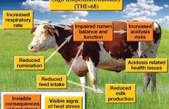
Clinical signs: Incubation period is typically 1–20 days. Most infections are noticeable after 3–7 days. Anthrax disease may be per acute, acute, sub-acute or chronic. The per acute form most often affects cattle, sheep and goats at the start of an outbreak and is characterized by staggering, trembling, difficulty in breathing, convulsions and death. Progression of the disease is rapid and premonitory signs may go unnoticed often animals are found dead with no rigormortis. Blood may fail to clot due to the toxin released by B. anthracis. The acute or subacute form is common in cattle, sheep and horses and manifest high fever, increased heart rate, excitement, depression, incoordination, cessation of rumination, low milk production, bloody discharges, respiratory distress, convulsion, and death within 48–72 hours. But the chronic form is usually seen in less susceptible species like swine.

Diagnosis: Careful microscopic examination of stained smears of blood, vesicular fluid, or edema may reveal the presence of B. anthracis. Biochemical and microbiologic tests provide a definitive diagnosis. It can also be isolated from skin lesions or respiratory secretions.
Treatment: Treatment of per acute case is usually untimely because of sudden death. Good recovery can be achieved through the use of anthrax antiserum in the early stage of the disease. Oxytetracycline at the dose rate of 5miligram/kilogram body weight parenterally can be effective if used from anthrax is most often seen in less susceptible species such as swine, but it has also been reported as developing in cattle, horses, dogs and cats. Route of infection in animals is most often ingestion, rather than inhalation or inoculation via skin lesions, initial suspicions of anthrax may be raised when livestock are found dead, bloated the onset of the disease. Penicillin streptomycin (Penstrep) also found effective if given concurrently with antiserum for 5 consecutive days.
Prevention: Anthrax can be prevented through annual vaccination programs. Rapid detection and reporting, quarantine, treatment of sub clinically affected animals (post exposure prophylaxis) and burning or burial of suspect and confirmed cases may prevent spore formation.
3.2 Brucellosis
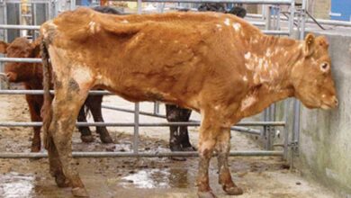
Brucellosis is a bacterial infection that can affect goats and other livestock such as sheep and cows and wild ruminants such as deer, elk and bison. Brucellosis causes abortion or stillbirth in animals. Brucellosis is one of the widest spread zoonosis transmitted by animals and in endemic areas, human brucellosis has serious public health consequences.
Etiology: Brucellosis in goats is normally caused by a Gram-negative coccobacillary rod, Brucella melitensis although Brucella abortus may also cause clinical brucellosis.
Clinical sign:Brucella melitensis is the most common cause of brucellosis in sheep and goats. It can cause abortion, retained placenta and swelling of the testicles. Abortion usually occur in late pregnancy in sheep and during the fourth month of pregnancy in goats. Communicably, brucellosis is contagious to humans. Bacteria are present in milk, placenta, fetal fluids, fetus, vaginal discharges, semen and urine. Ruminants and other animals can shed bacteria long-term or lifelong.

Diagnosis: History and clinical signs may be suggestive of the brucellosis. Demonstration of the bacteria in smears made from the samples of blood, bone marrow and other bod fluids can help confirming the diagnosis of the disease. Brucella can be isolated from the abomasal contents and lungs of the fetus, mammary glands, supramammary, retropharyngeal, parotid and mandibular lymph nodes and seminal vesicles by culturing on 5–10% blood or selective serum agar. Other serological methods like Serum Agglutination Test, Rose Bengal Plate Test, Enzyme Linked Immuno-Sorbent Assay (ELISA), Agar Gel Immuno-Diffusion (AGID) and Complement Fixation Test can be diagnostically used in the confirmation of brucellosis.
Treatment: There is no specific treatment of brucellosis that is successful, but long term antibiotics treatment can eliminate B. melitensis infections in valuable goats but the reproductive performance may be poor.
Prevention: Prompt vaccination of cattle, sheep and goats is recommended especially in endemic areas. Good hygiene practice such as milk pasteurization, proper meat processing, correct handling of stillbirths and animal carcasses are important strategy for the prevention of brucellosis in goats.
3.3 Caseous lymphadenitis (CLA)
Caseous lymphadenitis is an infectious disease caused by the bacterium Corynebacterium pseudotuberculosis, that affects the lymphatic system, resulting in abscesses in the lymph nodes and internal organ of Goats and Sheep.

Transmission: The disease is highly contagious that affects sheep and goats. When abscess ruptures, it releases a huge number of bacteria on to the skin and wool and it results to the consequent contamination of the surrounding environment. Animals may get infected when come in contact with the affected animals or indirectly via already contaminated fomites. Infected animals may contaminate feed and water, which may become source of infection. The disease is also easily spread through the materials that are used during the operation of the animals such as castration, identification with ear tags or by tattooing. It is thought to also be spread by coughing or even by flies.
Etiology: CLA is caused by a gram-positive, nonmotile pleomorphic rods bacterium known as Corynebacterium pseudotuberculosis that has a characteristic Chinese letter arrangement in the smear. When this organism successfully established in the host, it surrounds and subdue the immune system, as a result it causes chronic infection that may remain in the animal for life but not pestilent
Clinical signs: In CLA, there is abscessation in the region of peripheral lymph nodes especially the submandibular, parotid, prescapular and prefemoral nodes, which is termed superficial or cutaneous form. The internal form of CLA more commonly presents as dyspnea, loss of weight and failure to thrive. Other clinical signs include large pus filled cyst on the neck, sides and udders, cough, purulent nasal discharge, fever and tachypnea with abnormal lung sounds may be observed.
Diagnosis: A provisional diagnosis of the disease can be based on clinical features and physical examination of lesions associated with lymphnodes. Confirmation of the disease is achieved by bacterial culture of suspected lesions and purulent materials from an intact abscess. Serologic tests are available but their reliability is unrealistic.
Treatment: Treatment of CLA is often unsuccessful, but supportive care can be helpful. However, CLA abscesses must be treated to prevent ruptures and further contamination of other animals and environment. Parenteral antibiotics may be used in severe cases. Surgical drainage of the affected lymph nodes is recommended.
Prevention: The prevention of CLA can be achieved through strict biosecurity measures, immediate isolation of the affected animals from the flock. Surgical procedures such as castration, shearing or mass vaccination should be carried out through aseptic means and affected premises should be disinfected thoroughly.
If you have any questions or need further information, please feel free to contact us at professionaldvm129@gmail.com. We are always here to assist you.


cllohp
After a stressful week, the calm I found at 인천여성전용마사지 felt like a deep breath I didn’t know I needed.
Heya! I just wanted to ask if you ever have any trouble with
hackers? My last blog (wordpress) was hacked
and I ended up losing a few months of hard work due to no backup.
Do you have any methods to stop hackers?
Stop by my web page: official teslla338 (http://www.dotank.kr)